
Ear Reconstruction
Hi this is Whitney Lane and Hani Naga, Duke Plastic Surgery Residents on the Resident Review – a Duke Plastic Surgery Podcast. Today we are continuing our Quick Hits series designed to review high yield topics for our yearly inservice examination.
Today we are going to be discussing high yield topics related to ear reconstruction.
Anatomy:

Anterior: Outside -> Inside.
- Helix: incompletely folded outer rim of the ear
- Antihelix: marks the floor of the auricle, normally protrudes further out than helix
- Tragus, antitragus: small protrusions that cover the concha
- Concha: bowl leading into the ear canal
See diagram for more details.
Vascular anatomy:

Posterior auricular artery: anterior/posterior surface of auricle
Superficial temporal artery: lateral surface of auricle
Occipital artery: supplies posterior auricular skin
Veins: drain into external jugular
Nerve anatomy:
Great auricular nerve (C2/C3)—
- Innervation: lower half of the ear
- Anatomy: ascends from ERBS POINT
- ERBS POINT = 6.5 cm below the level of the tragus, midpoint of SCM, posterior to EJ
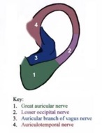
Lesser Occipital Nerve (C2/C3)
- Innervation: Posterior auricle
- Anatomy: higher than ERBS point and travels along the posterior border of the SCM
Auriculotemporal Nerve (V3)
- Innervation: supplies tragus, external auditory canal, anterior auricle
- Anatomy: ascends with STAs
Arnold’s Nerve: (auricular branch of vagus)
- Innervation: concha and posterior auditory canal
- Anatomy: travels along the ear canal
Lymphatic drainage:
Correlates with embryologic hillocks
- Tragus, root of helix, superior helix (from first branchial arch) – parotid nodes
- Antihelix, antitragus, lobule (second branchial arch) – cervical nodes
Aesthetic relationships of ear:
- The ear should be one ear length posterior to lateral orbital rim
- The superior edge to superior edge to inferior edge; 5.5-6.5cm,
- 2:1 ratio of height to width
- Ear protrudes 1-2cm,
- The incline of the ear on average is 25 degrees and it tilts 20 degrees
Whitney:
Embryology:
External ear development:
- Quick review: The 6 brachial arches develop during the 4th week of gestation and branchial clefts (external) and pharyngeal pouches (internal)
- Development of the ear starts at 4 weeks and is completed at 20 weeks gestation
- External ear develops from the 1st and 2nd arches. These develop into the 6 hillock that fuse to form the ear.
- 1st arch — develops into anterior hillock (1-3) which forms the tragus, root of the helix, superior helix, malleus, incus
- 2nd arch – develops into the posterior hillocks (4-6) posterior helix, antihelix, antitragus, lobule, stapes
- The internal ear develops from grooves and pouches
- 1st brachial groove/cleft: External acoustic meatus
- 1st paryngeal pouch: Middle ear and eustatian tube
Aesthetic relationships of ear:
- The ear should be one ear length posterior to lateral orbital rim
- The superior edge to superior edge to inferior edge; 5.5-6.5cm,
- 2:1 ratio of height to width
- Ear protrudes 1-2cm,
- The incline of the ear on average is 25 degrees and it tilts 20 degrees
Ear Anomalies:
Microtia:
- Definition: absence of external ear structures due to abnormal development of 1st, mandibular, or second- hyoid arches. The inner ear not affected
- Associated syndromes: Can be associated with Goldenhar or orbital auricular vertebral syndrome, 7th brachial clefts, and Treacher collins
- Treacher Collins is most associated with microtia due to abnormalities of the first and second branchial arches
- Treatment:
- Timing of ear reconstruction:
- Autologous reconstruction typically performed 6-7 years of age –> sufficient rib cartilage for reconstruction. Ear obtains 85% of growth at 3, fully developed by 5-7
- Timing of Hearing Aid Placement:
- For unilateral microtia: no need for BAHA (bone anchored hearing aid) placement prior to autologous reconstruction
- For bilateral microtia: external hearing aids can be placed 6-12 months, reconstruction 6-7yrs, ear canal creation 13-19.
- Type of Autologous Reconstruction:
- Nagata and Brent are the two most common techniques for autologous reconstruction. The difference between Nagata and Brent is in the number of stages (stages 2 vs 3 or more) and the reconstruction of the tragus and lobule
- Brent Reconstruction:
- Starts at age 6
- Requires 3 or more stages
- The tragus and lobule are elevated in separate procedures
- Nagata Reconstruction
- Starts at age 10 when there is more available cartilage
- Requires 2 stages
- The tragus and lobule are elevated in the same stage
- Other forms of reconstruction include:
- silastic frameworks not long lasting
- Alloplastic reconstruction can be successfully performed with the temporoparietal fascial flap to cover the implant (based on the superficial temporal artery)
Stahl ear:
Definition: consists of an extra cartilage fold (crus) – normally there are two an inferior and superior – in the scapha portion of the ear that can lead to unfurling of the superior helix giving the ear a pointed appearance
Treatment:
- Early: ear molding
- Late: two major schools of thought advancement of the third crus (IE local cartilage flap) to support the superior helix vs. excision of the third crus
Cryptotia hidden ear:
Definition: congenital deformity of cartilage of the scapha and antihelix– upper pole of ear is buried beneath scalp, superior auriculocephalic sulcus is absent
Cause: From abnormal distribution of the intrinsic transverse and oblique auricular muscles
Treatment: treatment is surgical release when child is older –>Helical release with STSG (superior portion of auricle)
Constricted Ear Deformities:
Definition: constricted/lop and cup ear deformities are a variety of deformities where the helical rim is either folded over or tight. These deformities fall along a spectrum and are based on the degree of cartilaginous and skin deficiency.
- Lop Ear: mild form of constricted ear due to protrusion of the ear, folding of the superior helix due to insufficiency of the helical cartilage with near normal skin.
- Cup ear deformity: more severe form of constricted hear with hooding of scapha and helix, flattening of antihelix
Treatment:
- Early: ear molding
- Late: surgical reconstruction based on the amount of deficit of the cartilage vs. skin.
Prominent ear:
Definition: characterized by widening of the conchoscaphal angle, increased auriculocephalic distance loss of antihelical fold, described as conchal valgus
Treatment: there are several surgical techniques for fixing prominent ear
- Mustarde technique for prominent ear: placement of permanent sutures through cartilage and perichondrium on cranial portion of ear to bend helix posteriorly from scapha cartilage to conchal cartilage
- Stenstrom’s: anterior surface of antihelix bent and cartilage scored to create posterior roll
- Luckett: crescent shaped portion of skin and cartilage excised from length of antihelix–> edges sutured to recreate fold
- Furnas technique: sutures placed from concha to mastoid to diminish size of concha. Inadequate reduction of concha can protrude lobule, overtightening of mastoid sutures lead to pinned back appearance
- Webster: corrects prominent helical tail by fixation of helical tail to concha
Complications:
- Most common is recurrence
- Telephone ear: excessive reduction of concha, inadequate correction of prominent upper/lower poles during otoplasty leading to a pinched middle appearance of the ear with excessive show of the upper and lower portions of the ear.
Ear Molding:
- Ear molding is a technique of reconstructing or fixing congenital ear abnormalities. It is effective because during the early post-natal perior maternal estrogens keep the cartilage pliable. Can treat prominent ear, cryptotia, lop ear, stahls ear.
- Start ear molding as early as 3 days and it can be initiated as far out as 3 months. However, When treatment is delayed several weeks success rate drops to 50% (90% in some studies)
- Complications: most common is skin ulceration
Trauma to the Ear:
- Blunt trauma:
- Hematoma most common complication
- Hematoma treatment: evacuate with bolster dressing
- Hematoma complication: cauliflower ear (subperichondrial hematoma over devascularized cartilage)
- Burns:
- Treatment: mafenide acetate, non-compressive dressings (pseudomonas most common cause suppurative eschar)
- Partial Ear Defects:
- Etiology: trauma vs. cancer excision – squamous cell cancer most common on ear
- Upper Third/Middle Third
- Partial Defect with perichondrium intact: contralateral skin graft vs. local skin flap
- Small <2cm full thickness defect: primary closure vs helical advancement (Antia-Buch procedure)
- >2cm full thickness defect: contralateral conchal cartilage flap covered with retroauricular flap vs. rib cartilage graft covered by a retroauricular flap vs. composite chondrocutaneous flap
- For large middle third defects you can also use: Chondrocutaneous rotation flap which is inferiorly based on antihelix, antitragus, or lobule and can be used to reconstruct up to 5cm defects.
** Retroauricular flaps are based on the posterior auricular artery and vein and the flap wraps around the conchal graft. This requires division of the flap in a second stage
- Lower Third Defects:
- Two stage retroarticular flap to re-create the lobule
- Concha Bowl Defects: retroauricular flap vs. postauricular island flap (revolving door) good for conchal defects and can replace entire concha
- Amputation
- Replantation:
- Microsurgical replantation of ear:
- Arteries: dissection and reconnect the large arteries to ear. These enter at the posterior aspect of pinna (external carotid, anterior auricular branch of STA, branch of occipital artery)
- Vein: If no venous outflow can be found, ear should still be reattached with microvascular techniques, followed by leech therapy for venous congestion
- Replantation in Children: In children with large loss of ear, may perform composite graft when microsurgical reattachment is not an option. Auricular composite grafts used for alar reconstruction –> should be no larger than 1.5cm –> continue to observe if cyanotic with eschar (treat like skin graft)
- Complications of Replatation:
- Chondritis:
- Sx: pain, swelling tenderness
- Tx: IV antibiotics (surgical if suppurative!);
- Suppurative infection of a reconstructed ear: incision and drainage with drains first prior to removal cartilage
- Venous congestion for ear replantation can be managed non-operatively- use leeches; arterial insufficiency requires surgery
- Duskiness of graft s/p composite graft: requires HBO to stimulate angiogenesis. Leave the composite graft in place for 2 weeks to allow for demarcation.
- Reconstruction of an Amputated Ear when Replantation is not Possible:
- Autologous Reconstruction:
- Most often is based on the creation of a graft with rib cartilage
- Banking Ear Cartilage: it has been reported that ear cartilage can be stored in a pocked with in the abdominal tissue prior to reconstruction. However, cartilage can lose its strength and architectural detail –> can become warped or contracted
- Alloplastic Reconstruction: Porous polyethylene implants: cover with TP fascia –> if graft exposed (small area) can be allowed to granulate in with dressing changes, do not remove until 6 month post op
- Prosthesis: Most aesthetic outcomes for ear recon when entire ear absent- placement of osteointegrated screws and prosthetic device when TP flap unavailable
Miscellaneous:
- Chondrodermatitis nodularis chronica helicis: painful, chronic nodular or cystic ulcerative lesions seen in superior pole of helix –> mistaken as malignancy –> complete excision, biopsy underlying cartilage, and close
Images from
Prendergast P.M. (2013) Anatomy of the External Ear. In: Shiffman M. (eds) Advanced Cosmetic Otoplasty. Springer, Berlin, Heidelberg. https://doi.org/10.1007/978-3-642-35431-1_2